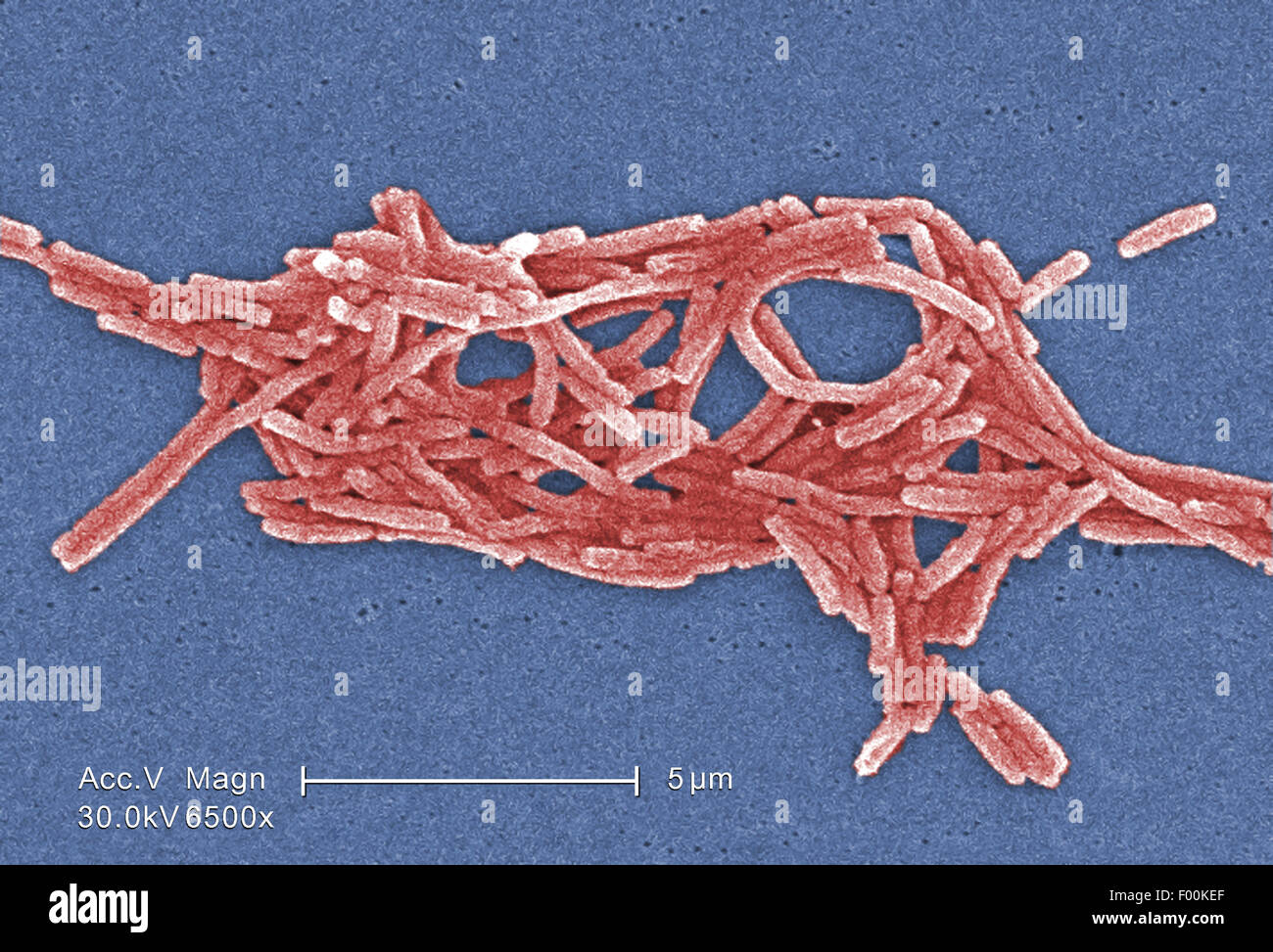
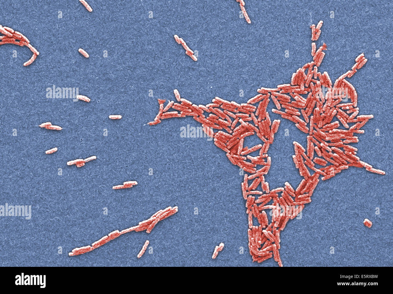
This scanning electron micrograph (SEM) depicted a number of red
$ 20.99 · 4.7 (660) · In stock

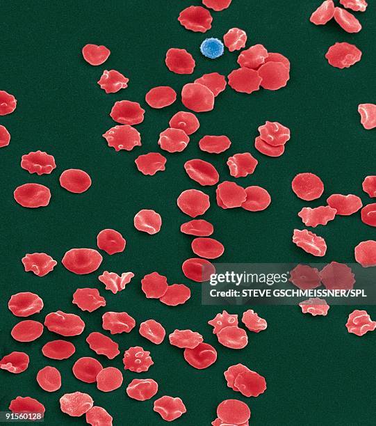
Download this stock image: This scanning electron micrograph (SEM) depicted a number of red blood cells found enmeshed in a fibrinous matrix on the luminal surface of an indwelling vascular catheter; Magnified 11432x Note the biconcave cytomorphologic shape of each erythrocyte, which increases the surface area of these hemoglobin-filled cells, thereby, promoting a greater degree of gas exchange, which is their primary function in an in vivo setting. In their adult phase, these cells possess no nucleus. What appears to be irregularly-shaped chunks of debris, are actually fibrin clumps, which when inside the living organi - 2BE0H0B from Alamy's library of millions of high resolution stock photos, illustrations and vectors.

Public Domain Picture, This scanning electron micrograph (SEM) depicted a closer view of a number of red blood cells found enmeshed in a fibrinous matrix on the lu, ID: 13546054811822
This scanning electron micrograph (SEM) depicted a number of red blood cells found enmeshed in a fibrinous matrix on the luminal surface of an indwelling vascular catheter; Magnified 2858x. Note the biconcave

Scanning electron microscopy hi-res stock photography and images - Page 3 - Alamy

Red Blood Cells, Sem #40 Duvet Cover by Science Source - Science

Solved The electron beam in a scanning electron microscope

This scanning electron micrograph (SEM) depicted numbers of
30 Biconcave Stock Photos, High-Res Pictures, and Images - Getty
Scanning electron microscopy bacteria hi-res stock photography and images - Page 3 - Alamy
Scanning electron microscopy (SEM) and transmission electron

Red Blood Cells, Rouleaux Formation, Sem #6 Poster by Science

Red Blood Cells, Sem #40 Framed Print by Science Source - Fine Art
Scanning electron microscope - Wikipedia