Figure 6 from Femoral Hernia: A Review of the Clinical Anatomy and
$ 15.99 · 4.6 (738) · In stock

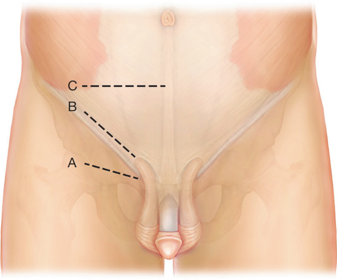
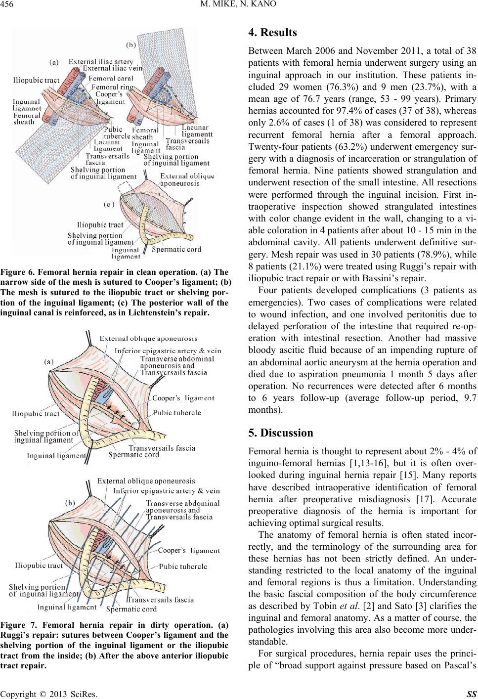
Figure 6. Femoral hernia repair in clean operation. (a) The narrow side of the mesh is sutured to Cooper’s ligament; (b) The mesh is sutured to the iliopubic tract or shelving portion of the inguinal ligament; (c) The posterior wall of the inguinal canal is reinforced, as in Lichtenstein’s repair. - "Femoral Hernia: A Review of the Clinical Anatomy and Surgical Treatment"

Femoral hernia, Radiology Reference Article
Femoral Hernia and Other Hidden Hernias: Options and Strategies
Femoral Hernia - A Review of Clinical Anatomy

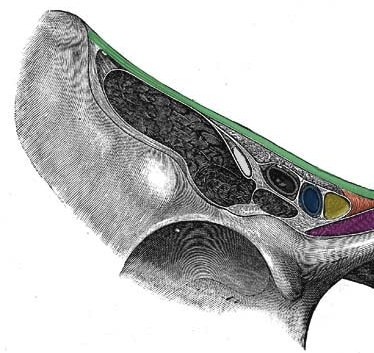
Intraperitoneal inspection. *: The inguinal ligament; L: Left; R

Hernias, Inguinal, Femoral, Umbilical
Femoral Hernia: A Review of the Clinical Anatomy and Surgical Treatment
Clinical Anatomy of the Groin: Posterior Laparoscopic Approach
![]()
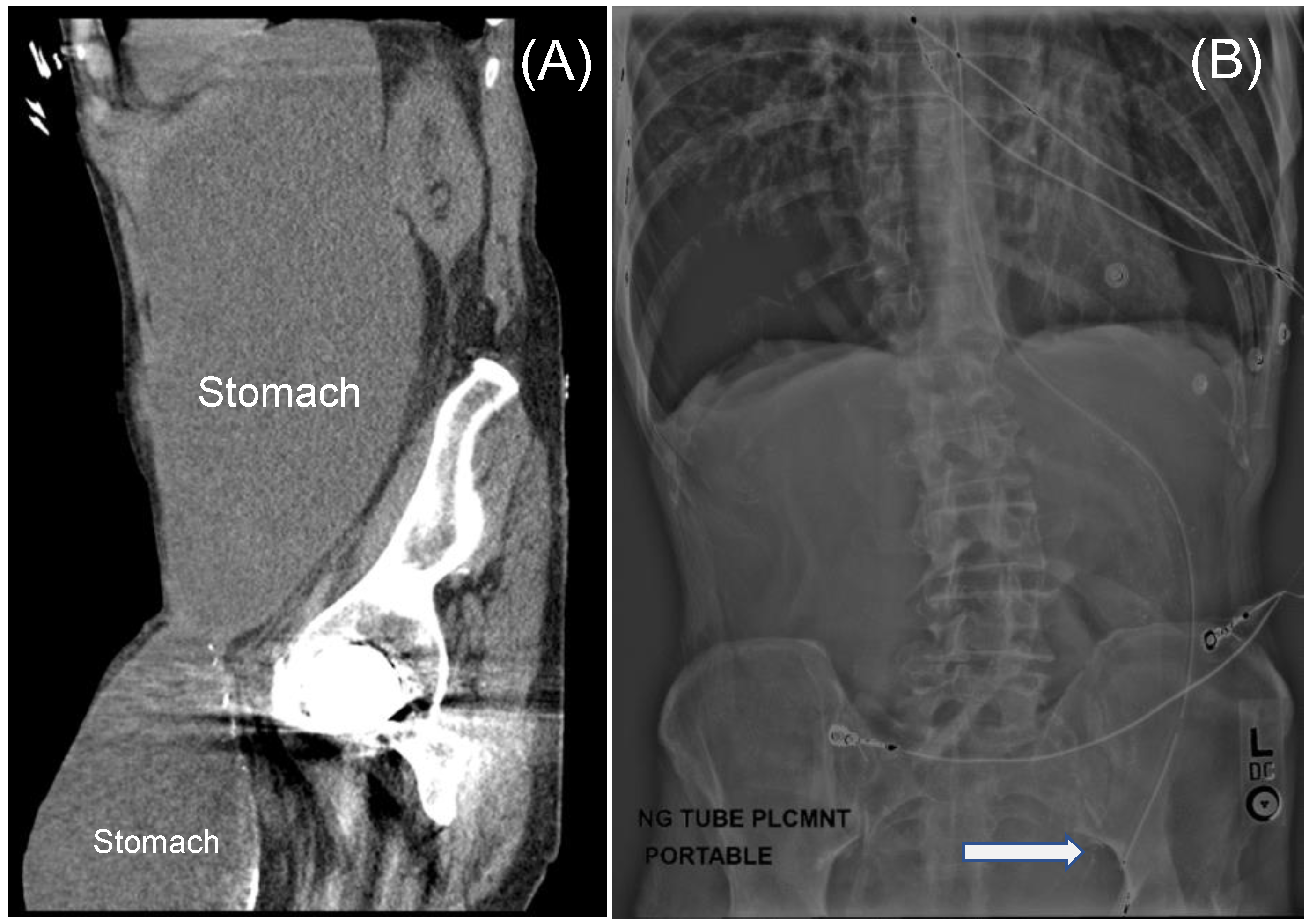
Left side femoral hernia – view from inside the abdomen (arrow)

Anterior and posterior views of myopectineal orifice ( from Elliott and

PDF] Laparoscopic repair of an incarcerated femoral hernia
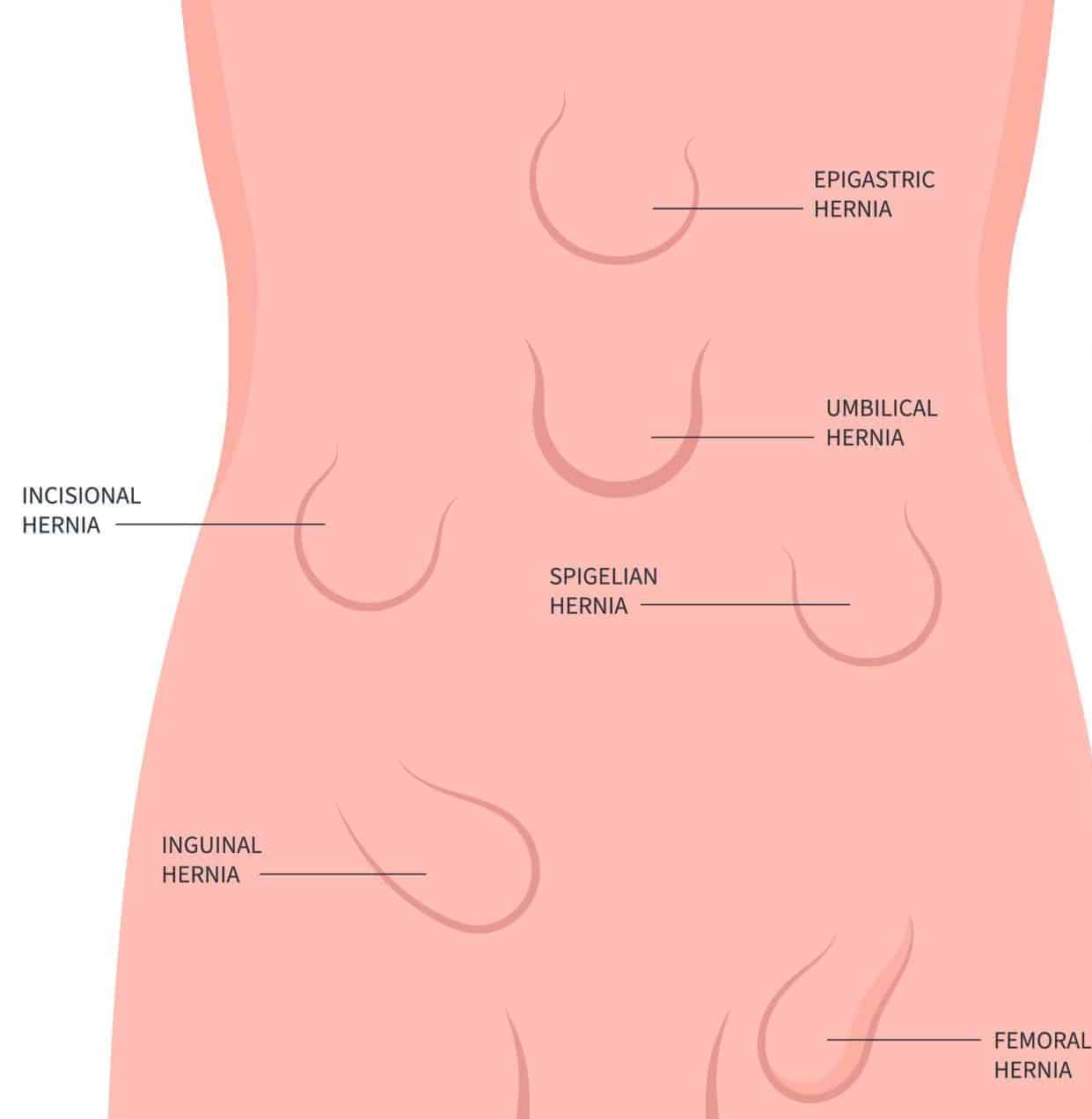
Abdominal Hernia - Epigastric - Spigelian - Obturator - TeachMeSurgery
JCM, Free Full-Text
Femoral Hernia - Risk Factors - Clinical Features - Management - TeachMeSurgery

An intraoperative image showing an erythematous appendix found within